|
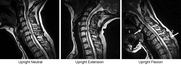
The upright-neutral image shows degenerative disc disease at multiple levels, a focal disc protrusion at C6/7 and narrowing of the central spinal canal (stenosis) at C6/7. Upright Extension demonstrates further central canal stenosis at C6/7. Upright Flexion reveals an anterior subluxation of C3 on C4 (arrow 1); also noted is hyperexpansion of the C6/7 interspinous space and laxity of the superspinous ligament indicating partial ligamentous rupture at this level (arrow 2). Finally, flexion also shows a reduction in central stenosis at C5/6 and C6/7. |