
Case courtesy of F.W. Smith, M.D. University of Aberdeen, United Kingdom
|
||||
| Previous<<1 2 3 4 5 6 7 8 9 10 11 12 13 14 15 16 17 18 19 20 >>Next | ||||
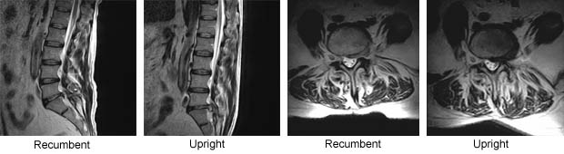
| Case#: 11 Hidden Nuclear Extrusion Uncovered by Dynamic Upright MRI | ||||
|
|
||||
| These scans show how recumbent only imaging can underestimate the maximum degree of pathology and not appreciate its dynamic nature. In the recumbent position you can clearly see the posterior prolapse at L5/S1. The upright, weight-bearing scan visualizes the extrusion of the nucleus pulposus through an annular tear into the spinal cord.This difference between recumbent and UPRIGHT® is visible in both the sagittal and axial views. | ||||
|
Case courtesy of F.W. Smith, M.D. University of Aberdeen, United Kingdom |
||||