|
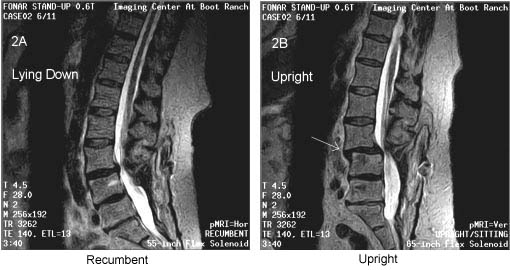
The patient was scanned in the FONAR UPRIGHT® MRI in early 2002, one year after her spinal fusion. Both Upright and recumbent scans were performed on her in the multi-position FONAR UPRIGHT® MRI. The recumbent MRI (left image) exhibited only a normal lumbar lordotic curve and a modest bulge of the L3-4 intervertebral disc, consistent with her prior recumbent MRI scan on a conventional MRI. The FONAR Upright scan (right image) revealed, however, a marked subluxation (anterolisthesis) at L3-4 and an accompanying spinal stenosis that were not visible on the recumbent MRI. |